

Members
Staff
Professor
Yoshito MASAMIZU, Ph.D.
E-mail: ymasamiz(at)mail.doshisha.ac.jp
ORCiD researchmap ResearchGate
Associate professor
Kaneyasu NISHIMURA, Ph.D.
E-mail: kannishi(at)mail.doshisha.ac.jp
ORCiD researchmap ResearchGateResearch
Neurons, once lost, cannot regenerate. However, if humans possessed the ability to regenerate lost neurons, some neurological diseases might have been overcome through our own healing capacity. How are the neurons that make up the brain formed, and how do they create networks with each other? By decoding this dynamic and complex process of neuronal formation and establishing technology to generate neurons, we believe we can contribute to maintaining people's health and to treating neurodegenerative diseases.
In the field of regenerative medicine, treatments to replenish lost neurons are being developed using cell transplantation with neural progenitor cells derived from pluripotent stem cells (human ES/iPS cells) and direct reprogramming that converts other brain cells directly into neurons. Using human iPS cells, we focus on Parkinson's disease and conduct fundamental research toward the functional and morphological regeneration of the lost midbrain dopaminergic neural circuits. We also develop novel therapeutic strategies from the perspective of drug discovery, reproducing molecular pathology of brain diseases by incorporating neural organoid preparation, genome editing, and imaging technologies.
Research Topics
- Induction of neurons and neural organoids by recapitulating human brain development
- Pathological modeling of neurodegenerative diseases and development of functional regenerative therapies
- Generation of neurons through manipulation of cell fate
Selected Publications
- Gima S., Oe K., Nishimura K.† et al., Host-to-graft propagation of inoculated α-synuclein into transplanted human induced pluripotent stem cell-derived midbrain dopaminergic neurons. Regen. Ther., 25, 229-237 (2024). †Corresponding author
- Nishimura K. et al., Single-cell transcriptomics reveals correct developmental dynamics and high-quality midbrain cell types by improved hESC differentiation. Stem Cell Reports, 18 (1), 337-353 (2023).
- Nishimura K.† et al., Rapid conversion of human induced pluripotent stem cells into dopaminergic neurons by inducible expression of two transcription factors. Stem cells Dev., 31 (11-12), 269-277 (2022). †Corresponding author
- Amimoto N.*, Nishimura K.*,† et al., Generation of striatal neurons from human induced pluripotent stem cells by controlling extrinsic signals with small molecules. Stem Cell Res., 55, 102486 (2021). *Co-first author,†Corresponding author
- La Manno G., Gyllborg D., Codeluppi S., Nishimura K. et al., Molecular diversity of midbrain development in mouse, human, and stem cells. Cell, 167 (2), 566-580 (2016).
- Nishimura K. et al., Estradiol facilitates functional integration of iPSC-derived dopaminergic neurons into striatal neuronal circuits via activation of integrin α5β1. Stem Cell Reports 6 (4), 511-524 (2016).
- Samata B., Doi D., Nishimura K. et al., Purification of functional human ES and iPSC-derived midbrain dopaminergic progenitors using LRTM1. Nat. Commun., 7, 13097 (2016).
- Nishimura K., et al., Identification of neurexophilin 3 as a novel supportive factor for survival of induced pluripotent stem cell-derived dopaminergic progenitors. Stem Cells Transl. Med., 4 (8), 932-944 (2015).
Associate Professor
Hironobu OSAKI, M.D., Ph.D.
E-mail: hosaki(at)mail.doshisha.ac.jp
ORCiD researchmap ResearchGateResearch Topic 1: How is "pain" generated in the brain? (Understanding pain neural circuits)
Pain is an important signal that tells us of bodily dysfunction, as many patients visit hospitals complaining of pain. However, since "pain" is a phenomenon generated in the brain, treating the affected area alone often cannot eliminate it. By understanding the neural circuits that generate pain, we aim to develop approaches for intractable pain conditions.
Related Publications
- We elucidated how pain sensation is processed in the primary somatosensory cortex.
Osaki# et al., Distinct nociception processing in the dysgranular and barrel regions of the mouse somatosensory cortex. Nature Communications (2022) #co-corresponding author - We elucidated how the thalamic reticular nucleus, an inhibitory nucleus in the sensory thalamus, selectively transmits specific signals to the cerebral cortex.
Osaki# et al., Receptive field properties of cat perigeniculate neurons correlate with excitatory and inhibitory connectivity to LGN relay neurons. Neuroscience Res. (2018) #corresponding author - (Collaborative research with Nihon University School of Dentistry, Dept. of Pharmacology) We provided a behavioral experimental system for verifying analgesic effects through neural activity control using quantitative pain evaluation.
Kobayashi, Osaki et al., Regulation of nociception by long-term potentiation of inhibitory postsynaptic currents from insular cortical parvalbumin-immunopositive neurons to pyramidal neurons. PAIN (2025)
Research Topic 2: Understanding neural circuit mechanisms contributing to recovery from brain injury
When brain neural circuits are damaged by stroke or other causes, the original function is lost, and various compensatory reactions occur in other brain regions. Capturing and understanding these changes is expected to provide knowledge useful for the development of neural circuit regeneration medicine. We also aim to develop methods that effectively harness such plasticity to further promote functional recovery.
Related Publications
- Osaki*#, Nakashima*, Iwai*, Masamizu#, Functional vulnerability of the M1/S1 transitional zone in post-stroke motor deficits in mice. Neuroscience Research (2026) *co-first author, #co-corresponding author
 Show detailsHide details
Rodent models of cerebral infarction serve as valuable experimental systems for elucidating recovery mechanisms and evaluating therapeutic efficacy. Because the brain's functional topography dictates the nature of deficits and the extent of recovery, precise localization of the infarction site is crucial for accurately assessing therapeutic effects.
In many previous studies, infarctions were induced and evaluated within specific anatomical regions, such as the primary motor cortex (M1) or the primary somatosensory cortex (S1). However, due to experimental limitations, infarctions frequently spanned both regions or exhibited significant spatial variability within each area. These limitations hinder the ability to establish a clear correlation between lesion topography and functional outcomes.
To address this, we developed a system to accurately map injury sites three-dimensionally onto a standard mouse brain template. This approach enables the longitudinal tracking of the relationship between the injury site and the severity of behavioral impairment.
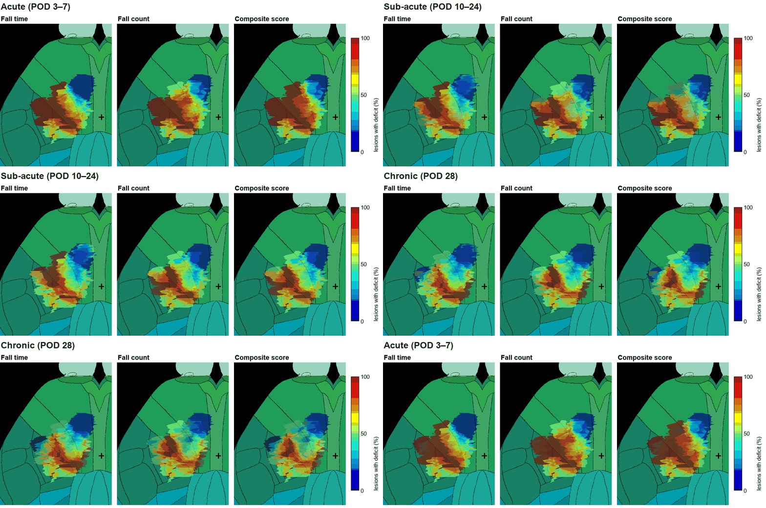
By systematically inducing infarctions in various locations, we found that damage to the transition zone between M1 and S1 results in behavioral impairments that tend to persist into the chronic phase. Conversely, when the infarction site is located outside this boundary, there is a tendency toward relatively early recovery, indicating that the transition zone between the motor and sensory cortices plays a crucial role in the recovery process. Furthermore, these results demonstrate the necessity of precise injury localization when evaluating therapeutic efficacy in cerebral infarction research. Consequently, these findings are poised to significantly influence evaluation methods in future studies.
Lesion-symptom maps over recovery (acute: POD 3–7 → sub-acute: POD 10–24 → chronic: POD 28). Redder pixels were covered mostly by lesions from the more-affected half of the cohort. The red territory contracts as the animals recover, while the region most associated with the deficit stays at the M1/S1 border (black cross: bregma).
Additionally, the program used in this study to register injury sites onto a standard brain template (the Allen Mouse Common Coordinate Framework (CCFv3)) and correlate them with behavioral data is available on GitHub, enabling its broad application to various cerebral infarction studies. This program can be applied not only to cerebral infarction sites but also to the three-dimensional identification and comparison of light stimulation sites using optogenetic methods.
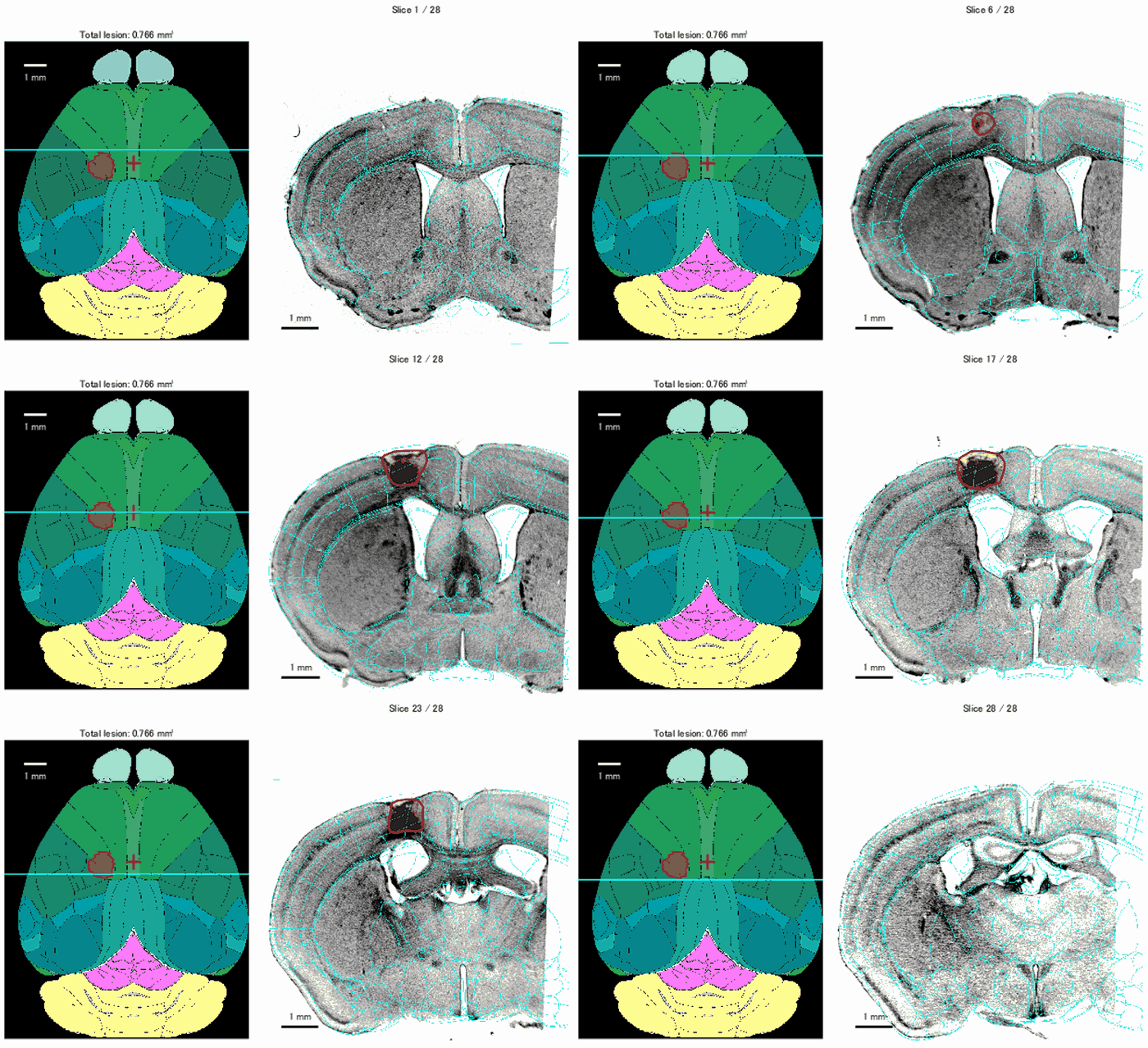
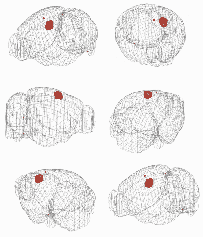
Example views from the program that registers lesions to the standard brain (CCFv3), for one animal. Left: the dorsal view with the projected lesion, and each coronal section registered to the atlas (cyan: atlas outlines; red: lesion outline). Right: the registered lesion in 3D inside the atlas brain outline.
Analysis code (GitHub): https://github.com/hironobu-osaki/lesion-symptom-map-leverpull
- Fukui*, Osaki*# et al., Layer-specific sensory processing impairment in the primary somatosensory cortex after motor cortex infarction. Scientific Reports (2020) *co-first author, #co-corresponding author
- Ishii*, Osaki* et al., Role of the medial agranular cortex in unilateral spatial neglect. Experimental Brain Research (2024) *co-first author
- Ishii*, Osaki* et al., Ipsilesional spatial bias after a focal cerebral infarction in the medial agranular cortex: A mouse model of unilateral spatial neglect. Behavioural brain research (2021) *co-first author
- Sato*, Itokazu*, Osaki* et al., Interhemispherically dynamic representation of an eye movement-related activity in mouse frontal cortex. eLife (2019) *co-first author
Methods
- Electrophysiology (OpenEphys)
- Ca2+imaging / intrinsic signal imaging
- Optogenetics (stimulus position scanning by galvano mirror control, etc.)
- Behavioral experimental systems combining electronics and programming

Research Specialist
Sachiko IWAI, M.S.
researchmapWith a background in applied biological sciences, I have gained extensive experience in a wide range of areas, from operating next-generation sequencers, cell culture, and molecular biology experiments to research on intractable eye diseases and drug discovery. I currently support neuroscience research as a research specialist.
Secretary
Yukie SEKIKAWA
Graduate student
D5 Daisuke NAKASHIMA
D3 Shintaro NUMATA
Alumni
Kotaro TEZUKA